Description
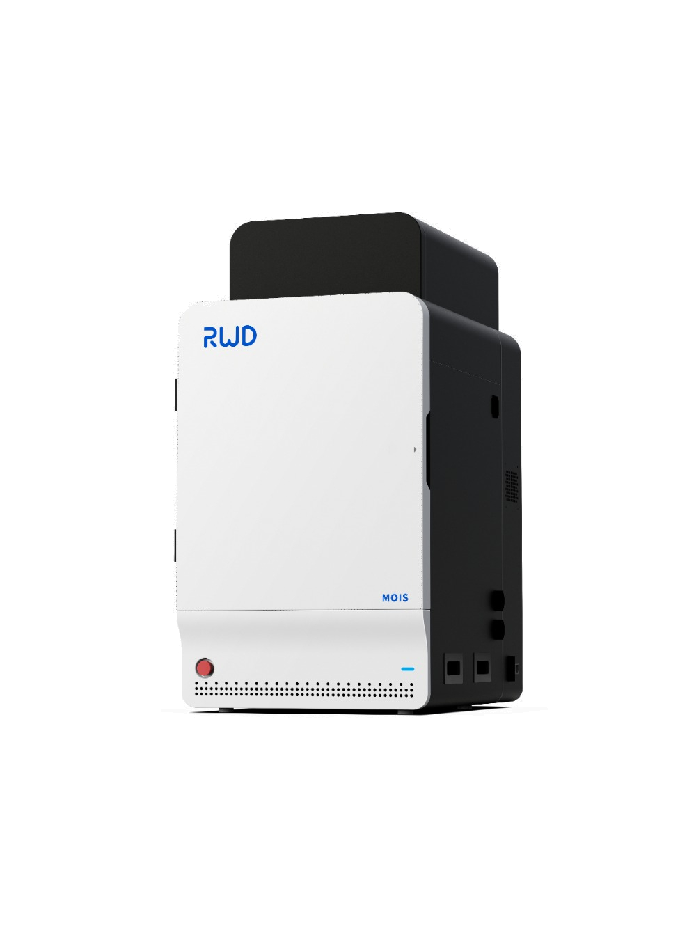
The Laser Speckle Imaging Contrast RFLSI-ZW takes advantage of LSCI technology and benefits from its countless advantages: contact-free, no contrast agents, high frame rate and high spatial resolution. They can be used to observe and record the blood perfusion of any exposed tissue or organ for studying microcirculation or for preclinical research such as ischemic stroke, lower limb, mesentery, etc. The system provides images and videos of blood perfusion (over 500 million pixels), quantitative data per unit of perfusion and vessel diameter.
From 2019, the RWD’s Laser Speckle Imaging Contrast has found wide use in more than 200 research institutes and universities around the world (Stanford University School of Medicine, Yale University, University of Manchester, Duke university, University College London, University of Tasmania, Universitaet Gesamthochschule Essen, Korea University, etc.) and contributed to the publication of as many articles and papers in renowned journals (Nature Neuroscience, Gut, Brain, Blood, Circulation Research, Nano Today, Nature Communications, Advanced Functional Materials, etc.).
Main areas of use:
- Monitoring of cerebral blood perfusion
- Evaluation of the MCAO model
- Observation of cortical spreading depression
- Research on hind limb ischemia
- Burning/transplantation of skin flaps
- Observation of organ microcirculation
- Skin allergies
- Septic shock
- Chicken chorioallantoic membrane test
- Analysis of limbs in the presence of diabetes


